This product has been viewed 4 times today.
Ordering in the UK? Contact us directly to request a quote and place an order sales@syngene.com
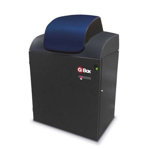
Looking for a Western blot imaging system that gives you more control over how you detect proteins? Look no further than the G:Box chemi XX6/XX9. This powerful system, powered by Syngene’s GeneSys software, lets you choose from a variety of chemiluminescence and fluorescence reagents from any manufacturer so that you can detect your proteins your way. With a 6 or 9 megapixel camera and HI-LED lighting options, you’ll get sensitive, publication-quality images with minimal background interference. Plus, the system has unlimited copies of GeneTools analysis software to help you maximise your data.
The G:Box XX6/9 is the perfect system for gel documentation, imaging and analysis in your lab. The in-house designed, FREE software with unlimited licences ensures you can detect chemiluminescence effectively and efficiently.
This system is perfect for imaging multiple sample types and sizes, from fluorescence 1D and 2D gels to chemiluminescent blots. Utilising the GeneSys automatic control software, with free updates, ensures your system remains on the cutting edge of technology, driven from a database containing hundreds of capture protocols. With a range of lighting and manual control options, image your way with the G:Box XX6/9
| SYSTEM | G:Box chemi XX6 | G:Box chemi XX9 |
| Image resolution (pixels m) | 6 | 9 |
| Effective resolution (pixels m) | 18 | 27 |
| A/D | 16 bit | 16 bit |
| Greyscales | 65,536 | 65,536 |
| Dynamic range OD | 4.8 | 4.8 |
| Quantum efficiency @ 425nm
|
73% | 73% |
| Lens (motor driven) | F0.80 or F0.95 | F0.80 or F0.95 |
| Stage | Moving | Moving |
| Filter wheel (7 position motor driven) | All fluorescence applications | All fluorescence applications |
| UV filter | Yes | Yes |
| Use with external PC | Yes | Yes |
|
DIMENSIONS |
||
| Max image area (cm) | 34.5×27.6 or 32.3×25.6 | 34.5×27.6 or 32.3×25.6 |
| Min image area (cm) | 15.6×12.5 or 15×11.8 | 15.6×12.5 or 15×11.8 |
| WxHxD (cm) | 57x99x55 | 57x99x55 |
| Weight (kg) | Approx. 45 | Approx. 45 |
| Power Input (V) | 100-240 | 100-240 |
|
LIGHTING |
||
| Epi LED White Lights | Yes | Yes |
| HiLED (Red, Blue, Green) | Optional | Optional |
| HiLED (Red, Infrared) | Optional | Optional |
| HiLED (Red, Blue, Green, Infrared) | Optional | Optional |
| HiLED (UV, Far Red) | Optional | Optional |
| Visible Light Converter | Optional | Optional |
| Blue Converter Screen | Optional | Optional |
| Slide-out UV Transilluminator 302nm (20cm x 20cm) | Optional | Optional |
| Edge Lighting Unit | Optional | Optional |
Our software is compatible with Windows 11*
*In-house testing has revealed no issues with the current version of Windows 11 (22H2 Update Version 22621.1702)